
Eye Associates Northwest — Advanced Imaging
Seeing Every Layer
of Your Eye Health.
From the front of the eye to the back of the retina, our imaging technology helps us diagnose and monitor eye disease earlier and more precisely.
- Corneal topography and Pentacam imaging
- Optical coherence tomography (OCT)
- Wide-field retinal photography with the Optos
Is This Right for You?
Am I a Candidate for Advanced Imaging?
Advanced imaging can help identify or monitor a range of eye conditions. If any of the following sound familiar, it may be time for an evaluation.
Imaging supports many diagnoses: Your doctor may recommend advanced imaging as part of a routine exam or in response to specific symptoms or risk factors, such as diabetes or a family history of eye disease.
The Technology
What Imaging Technology Do We Use?
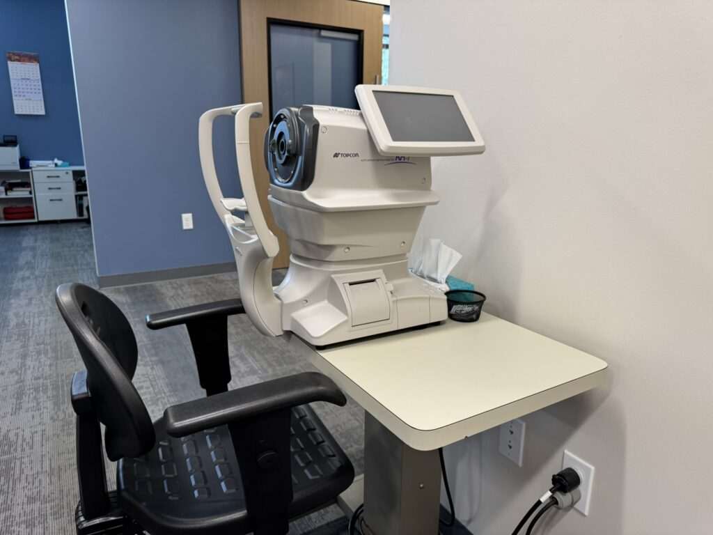
Eye Associates Northwest uses corneal topography and the Pentacam to assess the front surface of the cornea, supporting diagnosis of conditions such as keratoconus and helping determine candidacy for advanced intraocular lenses or laser refractive surgery. Optical coherence tomography (OCT) captures detailed cross-sections of the retina and evaluates the eye’s drainage angle for glaucoma risk. The Optos camera photographs the entire retina in a single wide-field image, and can be paired with fluorescein or indocyanine green dye injections to assess the retinal and choroidal blood vessels in detail.
Painless
All imaging tests are noninvasive and painless
No Downtime
Most patients resume normal activities immediately after imaging
Front to Back
Covers the cornea, retina, macula, and optic nerve
Same-Visit Review
Your ophthalmologist reviews imaging results with you during your appointment
Patient Reviews
Two Types of Imaging
Front-of-Eye vs. Back-of-Eye Imaging
Different imaging technologies assess different parts of the eye, and many patients benefit from both over the course of their care.
Corneal Imaging
Topography & Pentacam
- Assesses corneal shape and curvature
- Used for keratoconus and astigmatism management
- Helps prepare for advanced cataract or refractive surgery
- Does not assess the retina or optic nerve
Front of the Eye
Retinal Imaging
OCT & Optos
- Evaluates the retina, macula, and optic nerve
- Detects glaucoma-related changes and diabetic retinopathy
- Wide-field photography can document the entire retina
- Some tests may require pupil dilation
Back of the Eye
Your doctor will recommend the imaging that fits your symptoms, risk factors, or condition being monitored.
Step by Step
Your Imaging Visit
What to expect when advanced imaging is part of your appointment.
1
Check-In
- Technician reviews your medical history and symptoms
- Discussion of which imaging tests are recommended
2
Imaging
- Corneal topography, OCT, or Optos performed as needed
- Dye-based imaging (fluorescein or ICG) if indicated
3
Doctor Review
- Your ophthalmologist reviews images during your visit
- Findings discussed and compared to prior imaging if available
4
Follow-Up
- Repeat imaging scheduled to monitor any changes
- Treatment adjustments made based on imaging findings
What to Expect
Expected Advanced Imaging Outcomes & Recovery
Advanced imaging supports earlier detection and more precise monitoring of eye conditions over time.
What Patients Can Expect in the First 30 Days
- Most imaging tests require no downtime
- Temporary light sensitivity if pupils are dilated
- Bright flashes of light during OCT or Optos imaging are normal
- Vision may be blurry for a few hours if drops are used to dilate the eyes
Informed Care
Safety & Risk Considerations
Imaging itself carries minimal risk. Dye-based tests carry a small risk of allergic reaction, which your technician will screen for beforehand.
Safety Profile
- Noninvasive imaging for the cornea and retina
- Dye-based tests are administered under medical supervision
- Widely used across ophthalmology for decades
Possible Side Effects
- Temporary light sensitivity or blurriness after dilation
- Rare mild nausea with fluorescein dye injection
- Temporary yellowing of skin or urine after fluorescein dye, which resolves on its own
How We Minimize Risk
Trained Imaging Technicians
Our technicians are trained in the specific protocols for each imaging device used.
Physician Interpretation
Every image is reviewed by your ophthalmologist as part of your diagnosis or monitoring plan.
Screening Before Dye-Based Testing
Patients are screened for allergy history before fluorescein or ICG imaging.
Why Eye Associates Northwest
What Sets Our Imaging Program Apart
Comprehensive imaging coverage under one roof.
Full Range of Technology
From corneal topography to wide-field retinal photography, we cover the eye front to back.
Same-Visit Review
Your ophthalmologist reviews imaging results with you during your appointment whenever possible.
Disease-Specific Protocols
Imaging is tailored to the condition being evaluated, from diabetic eye disease to glaucoma.
Find Us
Advanced Imaging in Seattle & the Puget Sound
Advanced imaging is available at our Seattle-area locations.
Coverage & Financing
Cost & Insurance
Imaging is typically billed alongside your eye exam or as part of monitoring an existing condition.
Insurance Coverage
Diagnostic imaging tied to a medical condition, such as diabetes or glaucoma monitoring, is generally billed to medical insurance.
- Imaging performed as part of your comprehensive exam
- Separate charges may apply for specialized testing
- Billing support available through our office
- Discount available for qualifying self-pay patients
Eye Associates Northwest bills most major medical and vision insurance plans, including Medicare and Medicaid; check with your carrier about your specific coverage before your visit.
Self-Pay Options
Some imaging tests may fall outside your specific insurance benefits.
- Costs discussed before any specialized testing
- Multiple accepted payment methods
- Prompt-pay discount for qualifying patients
- Billing office available for questions
We accept cash, checks, Visa, MasterCard, American Express, and Discover, with a 15% prompt-pay discount for qualifying self-pay patients.
Insurance & Payment Made Simple. We bill most major medical and vision insurance plans, including Medicare and Medicaid. Patients without insurance coverage receive a 15% discount when paying in full the same day. Our billing team can answer questions about your specific coverage before your visit.
locationsCommon Questions
Frequently Asked Questions
What patients ask about our imaging technology.
Will imaging tests hurt?
No. Corneal topography, OCT, and Optos retinal photography are all painless, noninvasive tests performed in the office.
Do I need to be dilated for imaging?
Some tests, like certain OCT scans or fluorescein angiography, work best with dilated pupils, while others, like the Optos, can often be performed without dilation.
How often will I need imaging?
It depends on the condition being monitored. Patients with glaucoma, diabetes, or macular degeneration are often imaged at each visit to track subtle changes over time.
What is a fluorescein angiogram used for?
It uses a dye injected into the vein to highlight the retina’s blood vessels in detail, which helps assess vascular conditions like diabetic retinopathy and macular degeneration.
Ready When You Are
Schedule Your Free Consultation
Most patients see results within 24 hours. Consultations are available at all four locations, online scheduling takes less than two minutes.