
Eye Associates Northwest — Medical Retina
Focused Care
for the Back of the Eye.
The retina and macula are responsible for our sharpest, most central vision. We diagnose and treat the conditions that most commonly threaten them.
- Treatment for diabetic retinopathy and macular edema
- Management of dry and wet age-related macular degeneration
- Evaluation of new or worsening floaters
Is This Right for You?
Am I a Candidate for Medical Retina?
Several conditions can affect the retina and macula, often with distinct warning signs. If any of the following sound familiar, it may be time for an evaluation.
Sudden changes deserve prompt attention: A sudden worsening of floaters can signal posterior vitreous detachment or, in some cases, a retinal tear or detachment, which should be evaluated promptly.
The Anatomy
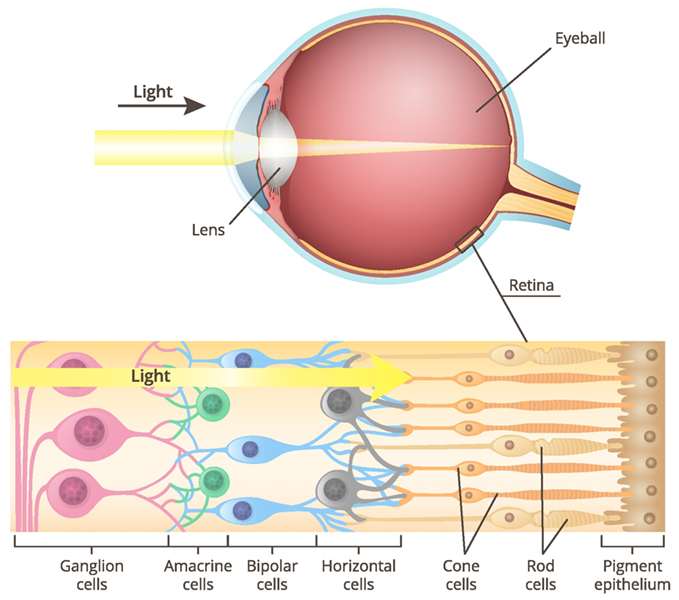
Anatomy of the Retina and Vitreous
The retina is the neurosensory tissue lining the back of the eye that receives light and relays visual information to the brain through the optic nerve. The macula, the central portion of the retina, is responsible for color perception and our sharpest central vision. The vitreous, a gel composed of collagen fibers and fluid, fills the central portion of the eye and plays a role in maintaining normal oxygen and nutrient balance after birth. Conditions affecting these structures include diabetic retinopathy, posterior vitreous detachment, and age-related macular degeneration, each requiring a different diagnostic and treatment approach.
Age-related macular degeneration is one of the most common conditions we treat, and one of the more complex to manage well. For a closer look at the differences between dry and wet AMD and how each is treated, visit our Age-Related Macular Degeneration page.
Dry & Wet AMD
Two forms of macular degeneration, each treated differently
Advanced Imaging
OCT and wide-field photography detect changes early
Injection Therapy
Available for wet AMD and diabetic macular edema
Ongoing Monitoring
Most retina conditions require monitoring even after treatment
Patient Reviews
Two Forms of AMD
Dry vs. Wet Macular Degeneration
Age-related macular degeneration comes in two forms, and treatment approaches differ significantly between them.
Dry AMD
Gradual thinning of the macula
- Occurs more slowly, typically with age
- Involves atrophy or thinning of the central macula
- Vitamin and antioxidant supplementation may slow progression
- Currently no treatment reverses existing damage
More Common Form
Wet AMD
Abnormal blood vessel growth
- Abnormal blood vessels leak or bleed into or under the retina
- Causes more rapid loss of central vision
- Treated with injections of medication into the eye
- Can sometimes slow or partially reverse vision loss with treatment
Less Common, More Urgent
A dilated retinal exam and imaging determine which form you have and the treatment plan that follows.
Step by Step
Your Medical Retina Journey
From evaluation to ongoing management of your retina condition.
1
Evaluation
- Dilated retinal exam
- OCT and, if needed, fluorescein or ICG angiography
2
Diagnosis
- Identification of the specific retinal condition
- Discussion of treatment urgency and options
3
Treatment
- Injection therapy, laser treatment, or vitrectomy surgery as indicated
- Nutritional guidance for dry AMD when appropriate
4
Ongoing Monitoring
- Scheduled follow-up imaging
- Treatment adjustments based on your response
What to Expect
Expected Medical Retina Outcomes & Recovery
Outcomes vary significantly by condition, and your retina specialist will set expectations specific to your diagnosis.
What Patients Can Expect in the First 30 Days
- Injection therapy often requires a series of treatments over time
- Some retina conditions require lifelong monitoring
- Vision changes between visits should be reported promptly
- Nutritional supplementation may be recommended for dry AMD
Informed Care
Safety & Risk Considerations
Retina treatments range from observation to injection therapy or surgery, each with its own risk profile that your specialist will discuss with you.
Safety Profile
- Injection therapy has an established safety record in treating wet AMD and diabetic eye disease
- Diagnostic imaging for the retina is noninvasive and painless
- Vitrectomy surgery is reserved for more advanced retinal conditions
Possible Side Effects
- Temporary eye irritation or redness after injection therapy
- Floaters or mild visual disturbance following certain treatments
- Risk of continued vision loss if advanced retinal disease is left untreated
How We Minimize Risk
Advanced Diagnostic Imaging
OCT and wide-field photography help detect retinal changes early, when treatment tends to be more effective.
Individualized Treatment Planning
Treatment is matched to your specific retinal diagnosis and its stage.
Close Monitoring
Regular follow-up imaging tracks your response to treatment and any need for adjustment.
Why Eye Associates Northwest
What Sets Our Medical Retina Program Apart
Focused expertise for conditions affecting the back of the eye.
Advanced Retinal Imaging
OCT and wide-field photography support early detection of subtle retinal changes.
Full Range of Treatments
From injections to laser treatment to vitrectomy surgery, we manage retina conditions at every stage.
Coordinated Diabetes and Eye Care
We work closely with patients managing diabetes to protect their retinal health.
Our Team
Our Medical Retina Team
Physicians experienced in diagnosing and treating diseases of the retina and vitreous.


Find Us
Medical Retina in Seattle & the Puget Sound
Medical retina consultations are available at our Seattle-area locations.
Coverage & Financing
Cost & Insurance
Medical retina evaluation and treatment are billed as medical care given the nature of these conditions.
Insurance Coverage
Retina conditions such as macular degeneration and diabetic retinopathy are medical diagnoses, so evaluation and treatment are typically billed to medical insurance.
- Comprehensive dilated retinal exam
- Advanced retinal imaging
- Injection therapy when indicated
- Billing support through our office
Eye Associates Northwest bills most major medical insurance plans, including Medicare and Medicaid; contact your carrier about coverage for injections or other retina treatments.
Self-Pay Options
For care outside your specific coverage, we keep the payment process simple.
- Pricing reviewed before treatment begins
- Multiple accepted payment methods
- Prompt-pay discount for qualifying patients
- Billing office available for questions
We accept cash, checks, Visa, MasterCard, American Express, and Discover, with a 15% prompt-pay discount for qualifying self-pay patients.
Insurance & Payment Made Simple. We bill most major medical and vision insurance plans, including Medicare and Medicaid. Patients without insurance coverage receive a 15% discount when paying in full the same day. Our billing team can answer questions about your specific coverage before your visit.
locationsCommon Questions
Frequently Asked Questions
Common questions about retina and macular conditions.
What causes macular degeneration?
Age-related macular degeneration comes in dry and wet forms. Dry AMD involves gradual thinning of the macula, while wet AMD results from abnormal blood vessels leaking or bleeding into the retina.
Are floaters always serious?
Not always, but a sudden increase in floaters can be a sign of posterior vitreous detachment or a retinal tear, so any sudden change should be evaluated promptly.
How is wet macular degeneration treated?
Wet AMD is generally treated with injections of medication into the eye to slow or, in some cases, partially reverse vision loss caused by abnormal blood vessel growth.
What is diabetic retinopathy?
It is damage to the retina’s blood vessels caused by chronically elevated blood sugar, which can be classified as nonproliferative or the more advanced proliferative form.
Ready When You Are
Schedule Your Free Consultation
Most patients see results within 24 hours. Consultations are available at all four locations, online scheduling takes less than two minutes.